


Severe internal diseases, poor nutrition and age slows nail growth and changes its structure. Only a doctor can accurately determine the cause of the disorder based on the results of tests and microscopic examinations.
But you can use the photo with different types of fungal diseases to get an idea of what is happening to the nails on the feet or hands.
Causes of nail deformation
Mold, yeast-like fungi and dermatophyte fungi cause infectious nail diseases (onychomycosis) with similar symptoms.
All kinds of nail or hand nail fungus deform the nail plate, change its transparency, gloss, color, this variety can be seen in the photos presented.
Changes in the nail occur not only with onychomycosis, but also with injuries, chronic paronychia (inflammation of the nail layer), psoriasis, eczema of the hands, dermatitis. Before deciding if you have a fungal infection, you should consider all possible options.
Signs of a fungal infection
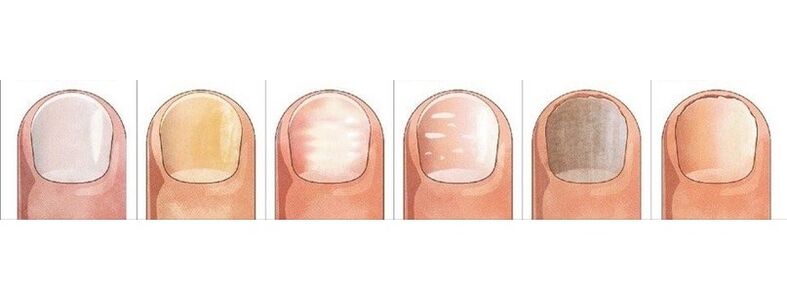
The most informative signs of fungal infection are changes in the color of the nail plate, the presence of nail shedding, surface changes - transverse, longitudinal grooves in the nail plate, dot deposits, thickening, destruction of the nail.
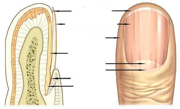
The pink color of a healthy nail is determined by the transparency of the nail plate and the blood vessels visible inside. With onychomycosis, the nail loses its transparency, the color becomes brown, yellow, less green, black.
Candida fungi and dermatophytes cause onycholysis - separation of the affected part of the nail. When infected with dermatophytes, onycholysis is observed on the far edge of the nail, and when infected with Candida, it remains behind the nail bed at the base of the nail, in the crescent part.
One of the symptoms of candidiasis may be inflammation of the lateral periungual ridges - paronychia. The disease has bacterial forms caused by streptococci and staphylococci, as well as non-infectious ones - eczema, psoriasis, systemic vasculitis.
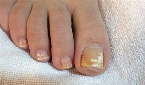
When toenails are affected by Trichophyton rubrum fungus, the plate is affected, as you can see in the photo, the roller is not affected by the infection. The plate turns yellowish, thickens strongly, and stands out well under the collected mushroom masses.
Nail fungus due to dermatophyte infection
In 95% of all nail fungi, the disease is caused by dermatophytes Trichophyton rubrum and Trichophyton mentagrophytes.
Trichophyton rubrum Infection
Onychomycosis begins when the fungus penetrates from the free edge to the bottom of the nail plate. Fungal infection is manifested by the appearance of a yellowish spot on the distal (distant) edge of the nail in the area of the stain, an uneven, scattered surface.
distal-lateral form of Trichophyton rubrum dermatophyte fungal infectionis common. In the photo, you can see that the stain caused by the application of the fungus is located along the lateral periungual nail fracture.
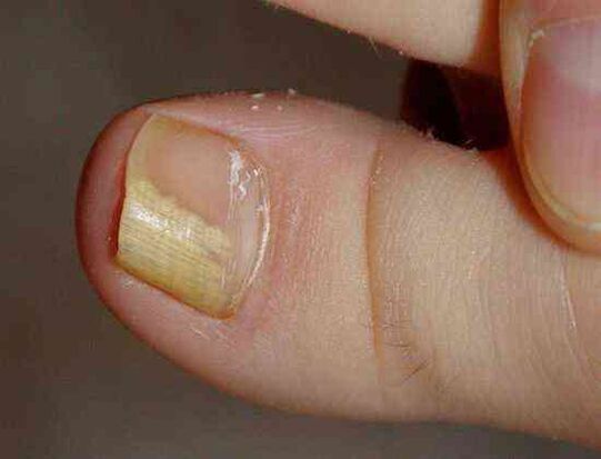
Trichophyton rubrum fungus usually causes hyperkeratosis by affecting the toes - the accumulation of fungus between the nail plate and the nail bed, which looks like an empty yellowish mass in the photo.
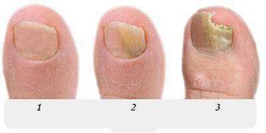
At this stage, the fungus, as in the photo presented, occupies an insignificant part of the nail, and with the help of local treatment it is possible to cope with the onset of onychomycosis.
Without treatment, the stain grows, gradually affecting the entire edge of the nail and then moving towards the crescent. In the photo, the nail fungus looks like yellowish streaks directed to the growth zone of the nail plate.
Withthe distal form of the nail fungus, which is often found on the thumbs,appears as a yellowish infection spot on the distal edge of the nail, in the center, as it were. seen in the photo.
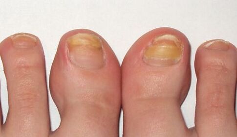
In the advanced stage of the fungus on the leg, as in the photo, several nails are affected, and treatment is no longer limited to local drugs and pills. In addition to antifungal medications, the nail apparatus is cleaned to completely or partially remove the nail plate.
As shown in the photo, hyperkeritosis should be treated with long-term therapy using all known antifungal agents and treatments caused by Trichophyton rubrum.
With general damage to the nail, the fungal infection spreads to the entire area of the nail plate, completely destroying the nail.
Infection with another member of the dermatophyte, Trichophyton mentagrophytes, can also cause a common fungal infection of the nail.Trichophyton mentagrophytes Infection
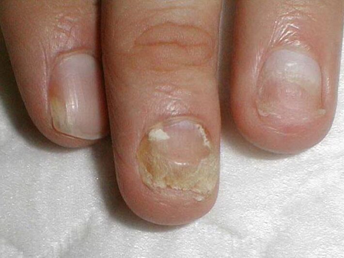
With the complete defeat of the nails by the fungus Trichophyton mentagrophytes, the nail plate is deformed, the photo shows that it thickens, changes its structure, collapses, yellowish spots appear on the entire surface.
Infection of the nail with this dermatophyte usually causes superficial white onychomycosis of the thumb, to a lesser extent the thumb.
This fungus practically does not occur on the nails of the hands, as in the photo, often causes digital dermatophytosis in the legs, and at the same time requires treatment of the skin of the feet and nails.
Symptoms of toenail fungus infection, usually on the feet, are white spots of various sizes, reminiscent of leukemia, as in the photo - the nail plate itself is a disease.
However, unlike leukonics, in which white spots are formed by the formation of air bubbles in the nail layers, white spots in fungal infections are the result of the activity of Trichophyton mentagrophytes.
Rarely, superficial white onychomycosis is caused by molds; The causative agent of this type of fungus in AIDS can be Trichophyton rubrum, which affects the nails of both feet and hands.
Changes in the nail due to Candida infection
The fungus usually occurs in women, mostly affecting the nails of the working hand in contact with water.
Proximal form of infection is characteristic of candidal onychomycosis, in which the fungus first affects the nail layer of the nail bed, then penetrates the growth zone and the nail bed. It then gradually moves along the nail from the edge of the nail and occupies a wider area of the nail plate.
The causative agent of candidal onychomycosis is Candida albicans. This fungus invades the feet and nails, extending from the crescent zone at the base of the nail plate to the free edge, as seen in the photo.
A symptom of Candida nail infectionalbicans is inflammation of the nail layer (paronychia), separation of the cuticle from the nail plate, pain, pus flow when the bacterial infection closes. .
Candida albicans can penetrate the nail and free edge. In this case, as a rule, they talk about a form of distal infection associated with candidiasis of the skin.
The treatment of candidiasis on the nails of the hands and feet, as shown in the photo, with damage to more than half of the nail plate, includes not only the fight against onychomycosis, but also measures to reduce the disease. activity in the natural reservoirs of the candidiasis - in the intestines, oral cavity, genital mucosa. . .
Infection with molds
Molds cause less fungus than Candida or dermatophytes. The main symptom of nail mold infection, as you can see in the photo, ischanging the color of the nail plate to blue, black, green.
Signs of nail mold can be dark spots, dots on the nail plate, or a long black stripe, as in the photo.
Preparations against fungi
Antifungal agents such as fluconazole, ketoconazole, terbinafine, itraconazole, grizeofulvin are used to treat nail fungus caused by dermatophytes, as in this photo.
Terbinafine antifungal agents are effective for dermatophyte infections.
Antifungal agents with voriconazole are highly active against dermatophytes.
Thisis used andnail moldis used on the feet, hands andagainst candidiasis. The spectrum of action includes molds such as Aspergillum, Fusarium, Penicillium.
Drugs based on itraconazole cope with molds.
Fungal nail diseases
sometimesappears withwith eczemaon thenail. In this case, the nail plate may move away from the nail bed, which is observed with a fungus.
Externally very similar to onychomycosismanifestations of psoriasis. With this disease, not onlydiscoloration, but alsonail plate thickens.
Dot deposits are found on the surface, the separation of the nail plate from the nail bed is noted. However, there are differences from fungi: in psoriasis, the separated and healthy parts of the toenail are separated over time by a pink, yellowish streak.
bluish colorgets a nailwith a false nail infection. Frequent mechanical friction of the nail layer causes the appearance of surface grooves and ripples of the nail.
Appearance is associated with metabolic disordersleukonychia white spotscan also be mistaken for a superficial white fungus with a large area of the stain. .
Changes in nail color and shape that cause damage. The toes are at the greatest risk. Like a fungus, a nail thickens and darkens with an injury.

The difference between an injury and a fungus is that the changes that occur during the injury are noted only on the injured finger, the nails of the other fingers remain unchanged, and the patient does not infect the finger, as in onychomycosis.
The result of trauma may be the partial separation of the nail from the nail bed, the formation of a cavity that is rapidly colonized by fungi under adverse conditions.
Nail plate, iron deficiency anemia, hormonal disorders can be separated from the nail bed under the influence of light (photonicolysis). Division, nail loss occurs with lichen erythematosus, bullous dermatoses, nail trauma.
But in the end, you can be sure that the result is correct and start treatment, you can only ask for help from a dermatologist - a dermatologist or a mycologist - a doctor who treats fungal diseases.